Tag: Quantum chemistry

  • Impossible molecules.

    Members of the chemical FAIR data community have just met in Orlando (with help from the NSF, the American National Science Foundation) to discuss how such data is progressing in chemistry. There are a lot of themes converging at the moment. Thus this article[cite]10.1039/c7np00064b[/cite] extolls the virtues of having raw NMR data available in natural product research, to which we added that such raw data should also be made FAIR (Findable, Accessible, Interoperable and Reusable) by virtue of adding rich metadata and then properly registering it so that it can be searched. These themes are combined in another article which made a recent appearance.[cite]10.1021/acsomega.8b03005[/cite]

    One of the speakers made a very persuasive case based in part on e.g. the following three molecules which are discussed in the first article[cite]10.1039/c7np00064b[/cite] (the compound numbers are taken from there). The question was posed at our meeting: why did the referees not query these structures? And the answer in part is to provide referees with access to the full/primary/raw NMR data (which almost invariably they currently do not have) to help them check on the peaks, the purity and indeed the assignments. I am sure tools that do this automatically from such supplied data by machines on a routine basis do exist in industry (and which is something FAIR is designed to enable). Perhaps there are open source versions available?

    17 18 19

     
    328[cite]10.1002/ejoc.201301308[/cite] 348 713

    Here I suggest a particularly simple and rapid “reality check” which I occasionally use myself. This is to compute the steric energy of the molecule using molecular mechanics. The mechanics method is basically a summation of simple terms such as the bond length, bond angle, torsion angle, a term which models non bonded repulsions, dispersion attractions and electrostatic contributions. The first three are close to zero for an unstrained molecule (by definition). The last three terms can be negative or positive, but unless the molecule is protein sized, they also do not depart far from zero. A suitable free tool that packages all this up is Avogadro.

    The procedure is as follows

    1. Start from the Chemdraw representation of the molecule. If the publishing authors have been FAIR, you might be able to acquire that from their deposited data. Otherwise, redraw it yourself and save as e.g. a molfile or Chemdraw .cdxml file.
    2. Drop into Avogadro, which will build a 3D model for you using stereochemical information present in the Chemdraw or Molfile.
    3. In the  E tool (at the top on the left of the Avogadro menu) select e.g. the MMFF94 force field. This is a good one to use for “organic” molecules for which the total steric energy for “normal” molecules is likely to be < 200 kJ. Calculate that for your system; this normally takes less than one minute to complete. The values obtained for the three above are shown in the table. All three are well over 200 kJ/mol, which should set alarm bells ringing.
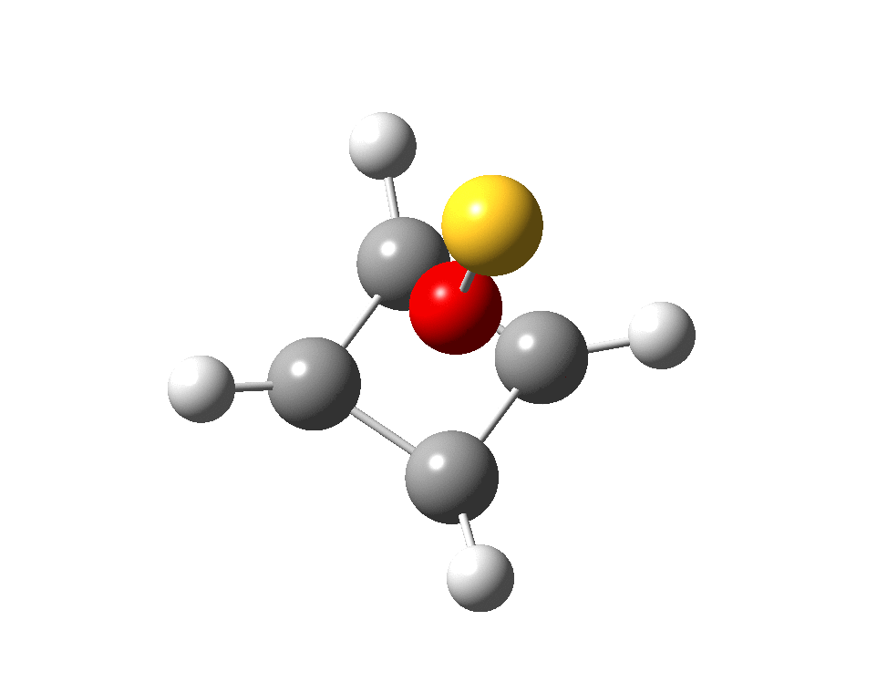
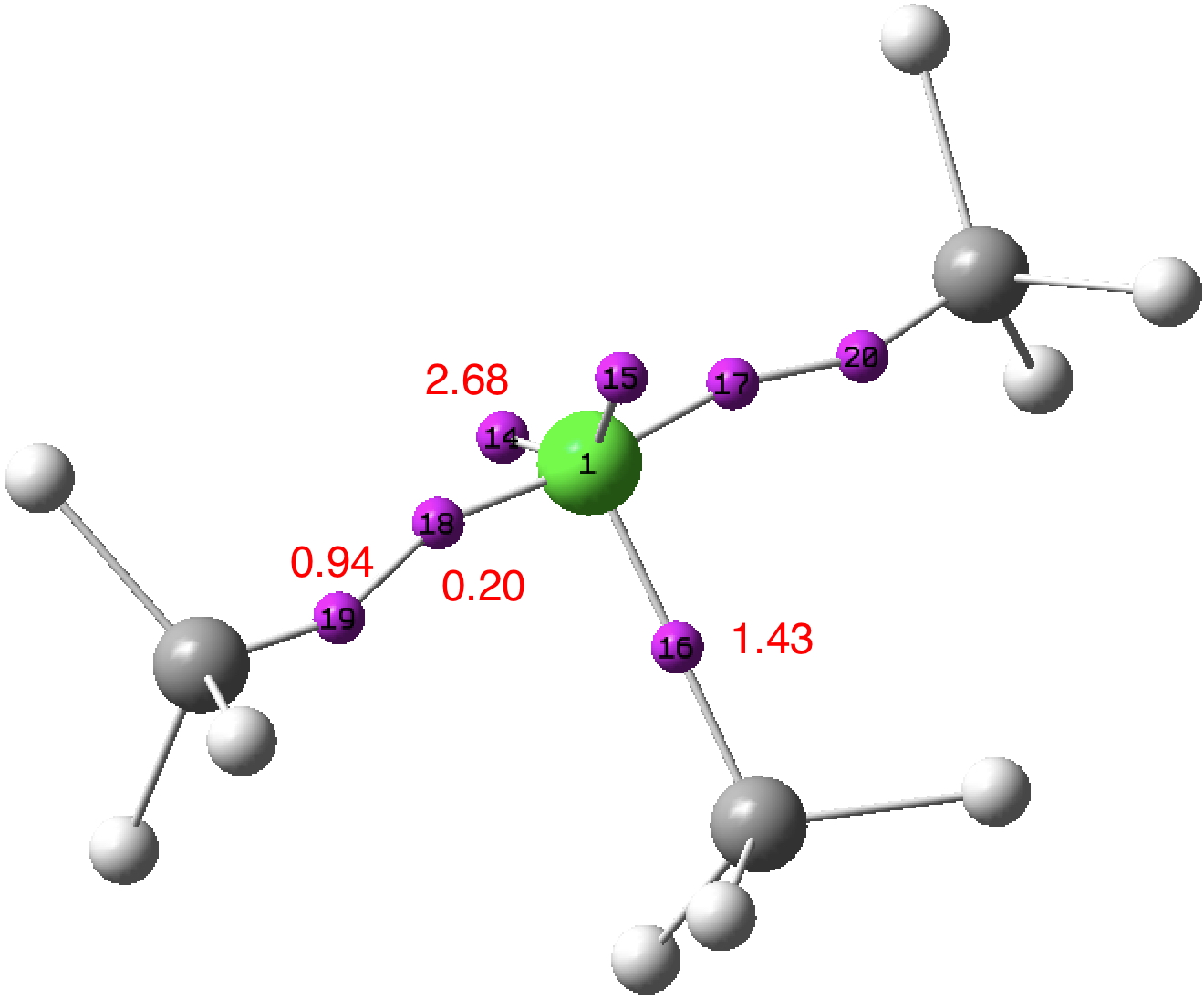
    4. A “more reasonable” structure for 17 is shown below. This has a steric energy of 152 kJ/mol, some 176 kJ/mol lower than the original structure. This does not of itself “prove” this alternative, but it is a starting point for showing it might be correct.Of course mis-assigned but otherwise reasonable structures are unlikely to be revealed by the steric energy test. But impossible ones will probably always be flagged as such using this procedure. 

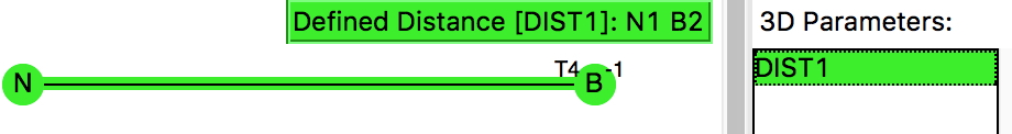
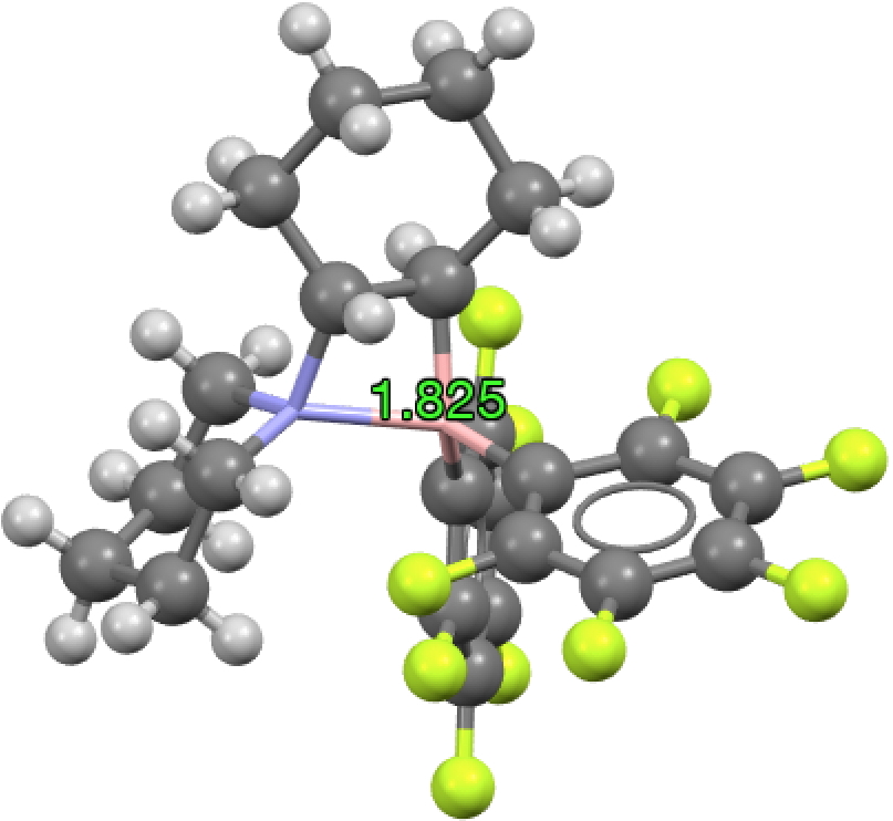
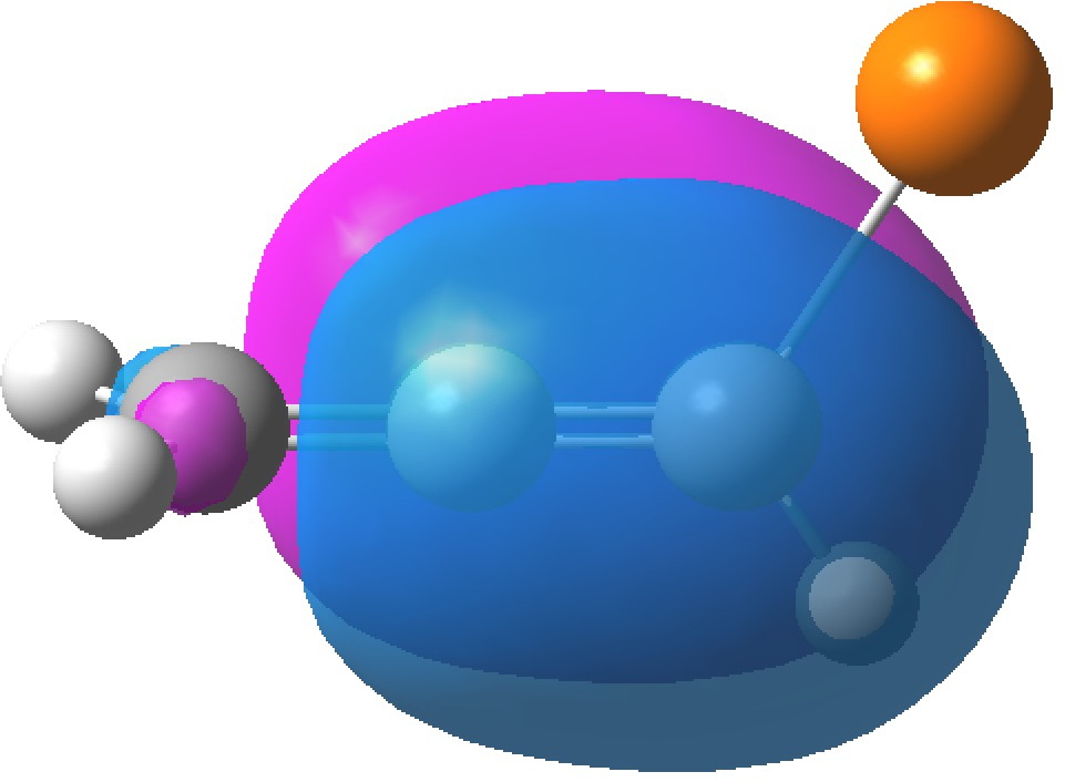
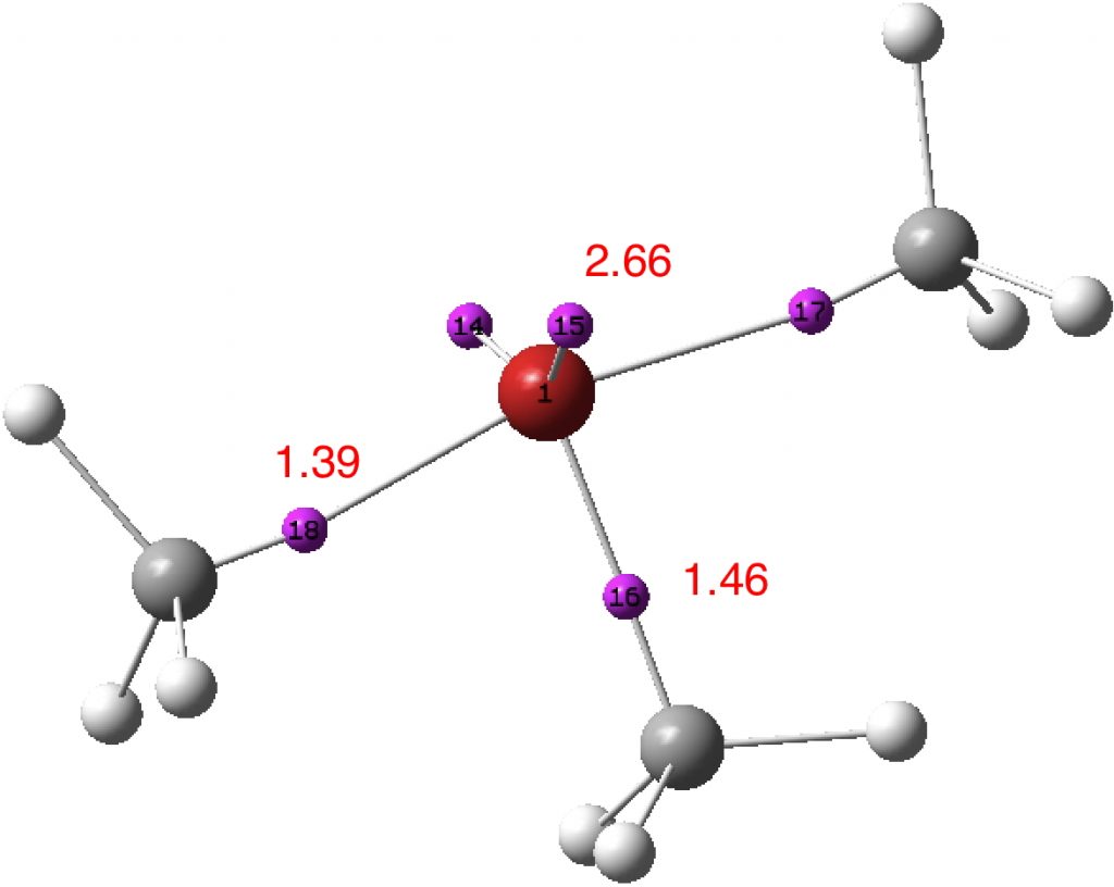
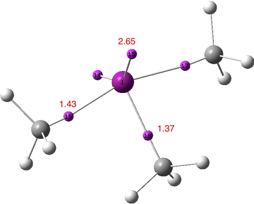
    Postscript: Hot on the heels of writing this, the molecule Populusone came to my attention.[cite]10.1021/acs.orglett.9b00423[/cite] On first sight, it seems to have some of the attributes of an “impossible molecule” (click on diagram below for 3D coordinates).

    However, it has been fully characterised by x-ray analysis! The steric energy using the method above comes out at 384 kJ/mol, which in the region of impossibility! This can be decomposed into the following components: bond stretch 30, bend 51, torsion 32, van der Waals (including repulsions) 177, electrostatics 87 (+ some minor cross terms). These are fairly evenly distributed, with internal steric repulsions clearly the largest contributor. The C=C double bond is hardly distorted however, which is in its favour. Clearly a natural product can indeed load up the unfavourable interactions, and this one must be close to the record of the most intrinsically unstable natural product known!

  • Why do flowers such as roses, peonies, dahlias, delphiniums (etc), exhibit so many shades of colours?

    It was about a year ago that I came across a profusion of colour in my local Park. Although colour in fact was the topic that sparked my interest in chemistry many years ago (the fantastic reds produced by diazocoupling reactions), I had never really tracked down the origin of colours in many flowers. It is of course a vast field. Here I take a look at just one class of molecule responsible for many flower colours, anthocyanidin, this being the sugar-free counterpart of the anthocyanins found in nature.

    These vary widely in the substituent around the aromatic rings, but here I take a look at just three differing substitutions. Thus pelargonidin has just one OH on ring C (R1‘, R3‘=H, see crystal structure[cite]10.3987/R-1985-10-2709[/cite]), cyanidin has two (R5‘=H, see crystal structure[cite]10.1107/S0567740877002702[/cite]) and is found in red roses, dahlia, peonies etc. Finally delphinidin (no crystal structure available) has three OHs in that region and is found in yes, delphiniums but also grape skins etc. Below is a colour table that allows one to relate the electronic transitions in a molecule to the observed colour, which of course is due to removal (absorption) of wavelength of light leaving us to see all the remaining wavelengths.
    colour table

    Next I show the computed UV/visible spectra of these three species (ωB97XD/6-311G(d,p)/SCRF=water). Click on any image to se a 3D model of the molecule.

    Note how in the visible region, all have a very simple (monochromatic) single electronic transition comprising mostly the HOMO→LUMO excitation.

    Click to view 3D model of the HOMO

    Click to view 3D model of the LUMO

    Now, λmax can be predicted quite poorly using most DFT methods, but the trends should be better predicted. Thus the change induced by adding two hydroxy groups is ~7nm, which is in effect how the colour seen in a flower can be tuned to display different shades.

    Next, pH. Using delphinidin, under basic conditions one can remove a proton from the cationic species to produce a neutral quinone. In fact, any one of five OH groups could have its proton removed and so it is of some interest to compare the relative energies of the five isomers so produced.

    Position proton removed Relative ΔG298, kcal/mol
    4′ 0.0
    5 3.8
    7 4.7
    3′ 11.8
    5′ 22.2

    In fact, one species only would have the major Boltzmann population (4′) and so we need only look at its UV/Visible predicted spectrum. This is shifted 17nm towards the red, thus producing a blue colour in what remains after it is absorbed. The absorption (ε) also increases significantly. Indeed the very striking colour of blue delphiniums (one of my favourite flowers) must be produced by such pH control in the plant. Given the presence of delphinidin in many grape skins, the next time I drink a glass of red wine, I will see if it turns blue upon adding some NaOH!


    FAIR data doi: 10.14469/hpc/4473

  • What are the highest bond indices for main group and transition group elements?

    A bond index (BI) approximately measures the totals of the bond orders at any given atom in a molecule. Here I ponder what the maximum values might be for elements with filled valence shells.

    Following Lewis in 1916[cite]10.1021/ja02261a002[/cite] who proposed that the full valence shell for main group elements should be 2 (for the first two elements) and 8 (the “octet“), Bohr (1922[cite]10.1007/BF01326955[/cite]), Langmuir (1919-1921[cite]10.1126/science.54.1386.59[/cite]) and Bury (1921[cite]10.1021/ja01440a023[/cite]) extended this rule to include 18 (the transition series) and 32 (the lanthanides and actinides). If we assume no contributions from higher Rydberg shells (thus 3s, 3p, 3d for carbon etc) and an electron pair model for orbital population (which amounts to the single-determinantal model), then the maximum bond index for hydrogen (and helium) would be 1, it would be 4 for main group elements, and then what?

    For the special case of hydrogen, I have previously identified (for a hypothetical species) a bond index of 1.33, due mostly to a high Rydberg occupancy of 1.19e. The more normal BI is <1.0, as noted for this hexacoordinated hydride system. My current estimate for the maximum bond index for main group elements is <4.5. Thus for SF6, it has the value of ~4.33 and that includes a modest occupancy of Rydberg shells of 0.36e = 0.18 BI. Exclude these and it is close to 4.

    Move on from group 16 to group 6 and you get compounds such as Me4CrCrMe44- or ReMe82- where the metal bond indices are ~6.5.‡ Compounds such as Cr(Me)6 (BI = 5.6)  and W(Me)6 (BI = 6.1) are rather lower. This is a long way from 18/2 = 9. The lanthanides and actinides[cite]10.1002/9781118688304.ch15[/cite] are unlikely to reveal many large BIs (32/2= 16 maximum value) since they are often ionic and the wavefunctions may be too complex to allow a simple index such as a BI to be safely computed.

    So if we are hunting for record BIs, the transition elements are the place to hunt. Can a BI of 6.5 be beaten? Can it even approach 9, its maximum value? Does anyone know of candidate molecules? 


    ‡FAIR Data doi: 10.14469/hpc/3352.

  • VSEPR Theory: Octet-busting or not with trimethyl chlorine, ClMe3.

    A few years back, I took a look at the valence-shell electron pair repulsion approach to the geometry of chlorine trifluoride, ClF3 using so-called ELF basins to locate centroids for both the covalent F-Cl bond electrons and the chlorine lone-pair electrons. Whereas the original VSEPR theory talks about five “electron pairs” totalling an octet-busting ten electrons surrounding chlorine, the electron density-based ELF approach located only ~6.8e surrounding the central chlorine and no “octet-busting”. The remaining electrons occupied fluorine lone pairs rather than the shared Cl-F regions. Here I take a look at ClMe3, as induced by the analysis of SeMe6.

    The difference between ClF3 and ClMe3 is that octet-excess electrons (two in this case) in the former can relocate into fluorine lone pairs by occupying in effect anti-bonding orbitals and hence end up not contributing to the central atom valence shell.‡ With ClMe3 the methyl groups cannot apparently sustain such lone pairs, at least not distinct from the Cl-C bond region. So might we get an octet-busting example with this molecule? A ClMe3 calculation (ωb97xd/6-311++g(d,p)) reveals a molecule with all real vibrational modes (i.e. a minimum, FAIR data DOI: 10.14469/hpc/3241) and ELF (FAIR data DOI 10.14469/hpc/3242)† basins as shown below:

    Density-derived approach: Two of the C-Cl bonds each exhibit two ELF basins; one disynaptic basin (0.94e) and one monosynaptic basin (0.20e) closer to the chlorine. The former pair integrate to 1.88e, density which largely arises from carbon (natural charge -0.84) and which contribute to a total integration for these carbons of 7.17e. The latter pair contributes to a total chlorine integration of 7.19e. The angle subtended at chlorine for the two 2.68e “lone pair” basins is 141°. Thus an inner, octet-compliant, valence-shell for chlorine is revealed, plus an expanded-octet outer one into which the two additional electrons go. The latter contribute to forming an octet-compliant carbon valence shell, but may be considered as not contributing to the valence shell of the other atom of the pair, the chlorine. An endo lone-pair rather than the more usual exo lone-pair if you will. These results reveal that the molecular feature we know as a (single) “bond” may in fact have more complex inner structures or zones, something we do not normally consider bonds as having. In this model, these zones are not invariably considered as shared between both the atoms comprising the bond.

    Orbital-derived approach: NBO analysis (FAIR data DOI: 10.14469/hpc/3241) reveals the chlorine electronic configuration as [core]3S(1.83)3p(4.67)4S(0.01)3d(0.03)5p(0.02,) showing very little population of the Rydberg shells (4s, 3d, 5p) occurs (0.13e in total). This method of partitioning the electrons allocates a chlorine Wiberg bond index of 2.00 and the methyl carbon bond index of  3.83. If the regular valence of Cl is taken as 1, then the central chlorine can be regarded as non-Rydberg hypervalent (the electrons in the 0.94e basins are taken as contributing to the chlorine bond index).

    The carbon-halogen bond internal structures simplify for Br (DOI: 10.14469/hpc/3248, 10.14469/hpc/3250) and I (DOI: 10.14469/hpc/3249, 10.14469/hpc/3247); for each only a single ELF basin is located and the NBO Br and I bond indices are respectively 2.10 and 2.1. This is not due to incursion of  Rydberg hypervalence (Br: [core]4S(1.83)4p(4.46)5S(0.02)4d(0.03)6p( 0.01); I: [core]5S(1.82)5p(4.29)6S(0.02)5d(0.02)6p(0.01) ) but of a merging of the carbon and halogen valence basin such that the ELF contributions to each cannot be deconvoluted. In each case the NBO bond indices of ~2 suggest hypervalency for the halogen.

    What have we learnt?  That the shared electron (covalent) bond can have complex internal features, such as two discrete basins for the apparently shared electrons. How one partitions these electrons can influence the value one obtains for the total shared electron count and hence whether the octet is retained or expanded for main group elements such as the halogens. And finally, that hypervalence and hyper-coordination are related in the orbital model at least. Thus along the series MenI (n= coordination number 1,3,5,7), the values of the Wiberg bond index at the halogen progress as 1.0, 2.1, 3.1 (DOI: 10.14469/hpc/3236) and 4.01 (DOI: 10.14469/hpc/3238), or one extra atom bond index per electron pair.  Given this, it seems useful to retain the distinction between the terms hypervalence and hyper-coordination, but also recognize that we still may have much to learn about the former.


    ‡See the previous post on SeMe6 for a more detailed discussion.

    † The FAIR Data accompanying this blog post is organised in a new way here. All the calculations are collected together with an over-arching DOI: 10.14469/hpc/3252 associated with this post, with individual entries accessible directly using the DOIs given above. The post itself has a  DOI: 10.14469/hpc/3255 and the two identifiers are associated with each-other via their respective metadata.  A set of standards (https://jats.nlm.nih.gov) with implementation guidelines for e.g. repositories, authors and publishers-editors  are expected in the future to establish infra-structures for cross-linking narratives/stories with the data on which they are based.

  • Elongating an N-B single bond is much easier than stretching a C-C single bond.

    An N-B single bond is iso-electronic to a C-C single bond, as per below. So here is a simple question: what form does the distribution of the lengths of these two bonds take, as obtained from crystal structures? 

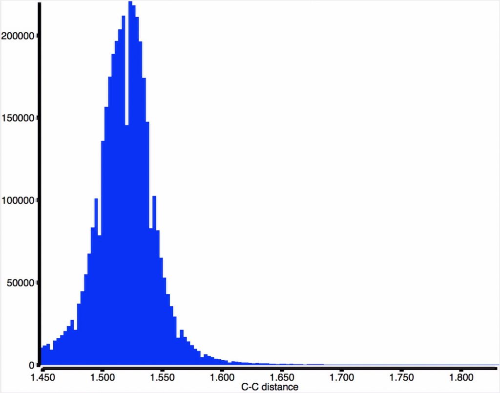
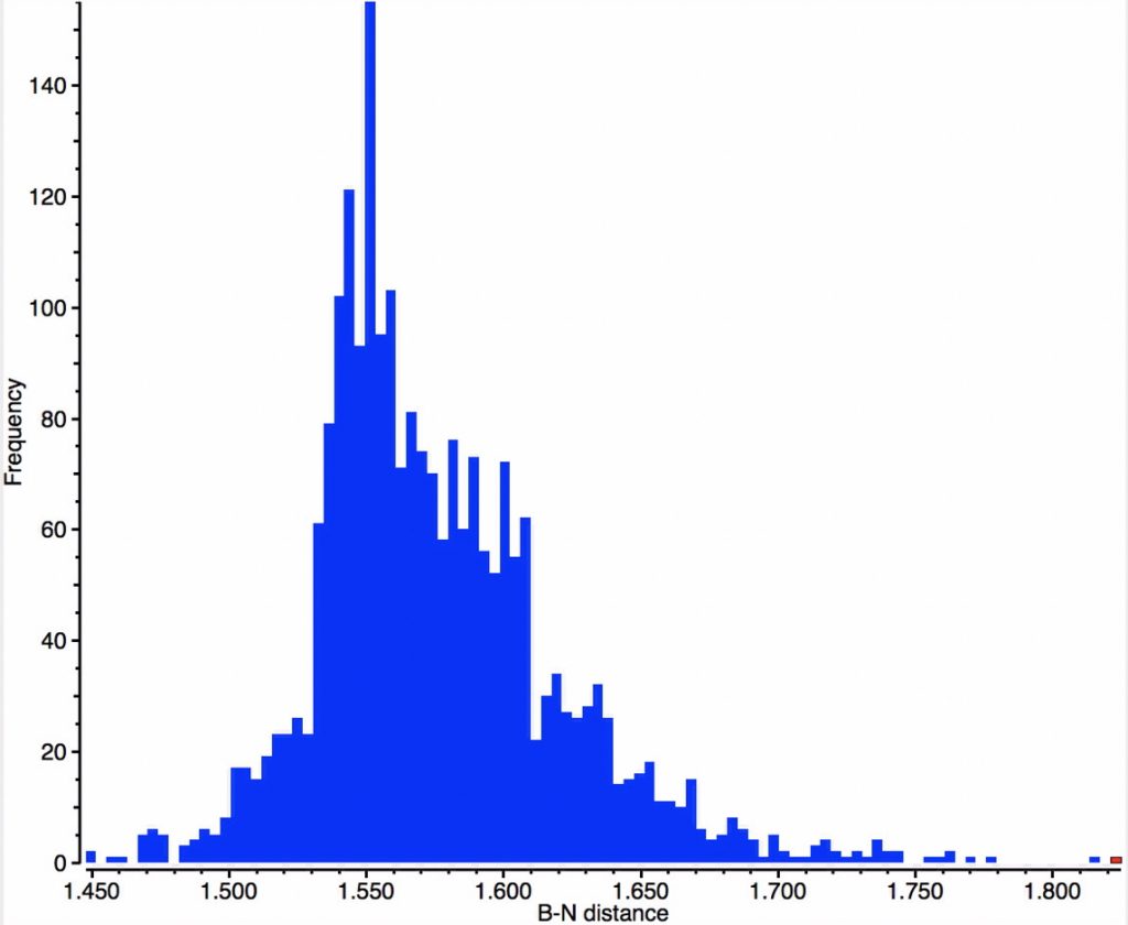
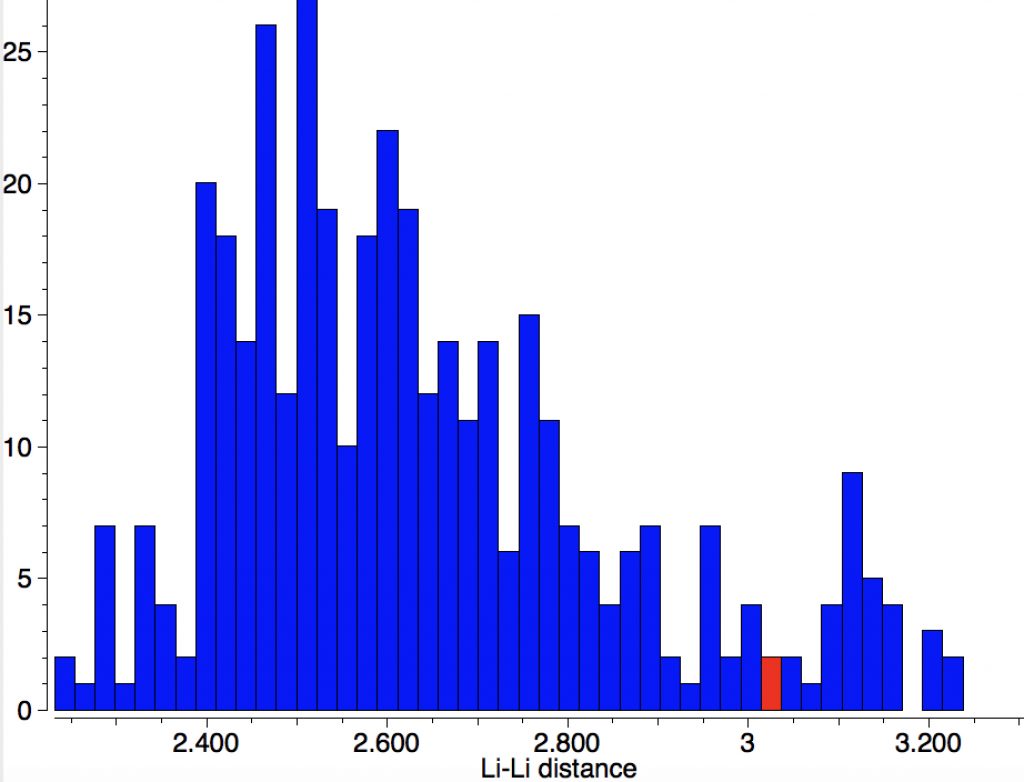
    The Conquest search query is very simple (no disorder, no errors).

    When applied to the Cambridge structure database (CSD) the following two distributions are obtained. That for carbon is pretty symmetric with the peak at ~1.53Ã… but with rather faster decay in the region >1.6Ã… compared with the region <1.46Ã… (the latter may be caused by hyperconjugation shortening the C-C bond).

    In contrast, the iso-electronic N-B distribution is more asymmetric about the peak of 1.56Ã…, exhibiting a long tail beyond 1.63Ã…, up to a value of 1.825Ã….

    The molecule with that longest N-B bond (1.825Å) is shown below; UWOHUK, Data DOI: 10.5517/ccwcwlp. This by the way is no crystal artefact; a calculation (ωB97XD/6-311G(d,p), Data DOI: 10.14469/hpc/3202) gives a calculated length of 1.81Å, with a N-B bond order of 0.48.

    Stretching a C-C bond heterolytically requires charge separation (a relatively unfavourable process) and likewise homolytic stretching would tend to form a biradical, in effect an excited state and again not favourable. In contrast, elongating the N-B bond reduces (at least formally) any charge separation and allows this heteronuclear pair to sustain (single) bond lengths over the much wider range of ~0.4Ã… without requiring biradical formation.

    One might wonder what other single-bonded atoms pairs give such unusually large spans in their bond length distributions.

  • The di-anion of dilithium (not the Star Trek variety): Another “Hyper-bond”?

    Early in 2011, I wrote about how the diatomic molecule Be2 might be persuaded to improve upon its normal unbound state (bond order ~zero) by a double electronic excitation to a strongly bound species. I yesterday updated this post with further suggestions and one of these inspired this follow-up.

    The standard molecular orbital diagram for Be2 below shows two electrons in both the 2s Σg and Σu levels, the first being considered bonding and the second antibonding. By exciting the two electrons from the Σu into the Πu MO to form a triplet, one converts one antibonding occupancy into two bonding occupancies, in the process changing the total formal bond order from zero to two.


    The triplet excited state of diberyllium

    You can see the results of my playing with these ideas both in my appended comments to the original post and the table below. This shows that the calculated bond order for the excited triplet state of Be2 is actually closer to 1.50 rather than to two, but definitely not zero!

    System Wiberg bond order Bond length FAIR Data
    Be2 singlet 0.15 2.805 10.14469/hpc/3082
    Be2 excited triplet 1.50 1.785 10.14469/hpc/3075
    Be22+ 1.00 2.135 10.14469/hpc/3076
    Be22- triplet 0.89 2.242 10.14469/hpc/3074
    Be22- excited singlet 3.00 1.817 10.14469/hpc/3083

    The games above represent isoelectronic substitutions and here I try one more, namely that Li22- is isoelectronic with Be2. Unlike the latter, there is no need to force an electronic excitation (ωB97XD/Def2-QZVPPD/SCRF=water) to achieve the required occupancies with Li22-.

    System Wiberg bond order Bond length FAIR Data
    Li22- triplet 1.501 2.381 10.14469/hpc/3087

    I also checked what crystal structures could tell us about Li-Li bonds and it seems 2.38Ã… is about as short as they get.

    At this point, the NBO analysis of the Li22- localised orbitals alerted me to another feature, which is that the Rydberg occupancy amounted to 2.18e. This in turn reminded me of the previous post which dealt with such occupancy in another small molecule, CH3F2-, but here the Rydberg occupancy involved the 3s/3p AOs of the carbon and the fluorine. With Li22- triplet, it is of the lithium 2p AO (2.18e) and only a tiny occupancy of 3d (0.03). By definition, for alkali metals such as Li the normal valence shell is just 2s, whereas 2p occupancy is considered a Rydberg state; a hypervalent state if you will. So Li22- triplet has a Li-Li hyper-bond!‡ Of course, by this definition most Li compounds are then hypervalent, since many have populated 2p shells.

    Even if use of the term hyper-bond to describe Li22- triplet is rather artificial, this example does reveal the games one can play with the first row elements Li-B (see table above). Given that most introductory text books on bonding normally only explain the diatomics formed from N-Ne (occasionally including C), I might suggest that these earlier elements are equally instructive and fun to play with.


    ‡ This species is 36.0 kcal/mol higher in free energy than two separated Li anions.

  • First, hexacoordinate carbon – now pentacoordinate oxygen?

    The previous post demonstrated the simple iso-electronic progression from six-coordinate carbon to five coordinate nitrogen. Here, a further progression to oxygen is investigated computationally.

    The systems are formally constructed from a cyclobutadienyl di-anion and firstly the HO5+ cation, giving a tri-cationic complex. There are no examples of the resulting motif in the Cambridge structure database. A ωB97XD/Def2-TZVPP calculation (DOI: 10.14469/hpc/2350) shows it is again a stable minimum, with a Kekule mode of 1203 cm-1.

    A QTAIM  topological analysis of the electron density shows it differs from the nitrogen analogue in now having the ring topological feature for the basal four carbons, which in turn gives rise to a cage critical point (blue dot). The values of the electron density are lower than for N.

    The ELF basin analysis shows the C-C bonds are regular single ones (2.01e), whereas the C-O bonds have a slightly greater electron population than the C-N bonds discussed in the previous post.

    I suspect the prospects of making a stable tri-cation in such a small molecule are lower than the crystal di-cation achieved with carbon as the apical atom. But the charge can be reduced to a di-cation by replacing the HO5+  above with S-O5+; the animation below showing the Kekule mode (1140 cm-1, DOI: 10.14469/hpc/2356).

    And for some (negative) loose ends.

    1. The P equivalent constructed from cyclobutadienyl di-anion and HP4+ is now unremarkably 5-coordinate. But in fact it is not a stable minimum (DOI: 10.14469/hpc/2357), having two negative force constants.
    2. as does the system  from cyclobutadienyl di-anion and O=P4+(DOI: 10.14469/hpc/2358)
    3. and the system from cyclobutadienyl di-anion and HS5+(DOI: 10.14469/hpc/2360).
    4. Transposition of S/O to give O-S5+ likewise (DOI: 10.14469/hpc/2359).

    So the family of hyper-coordinate 2nd row main group elements now comprises the experimentally verified C, with N and O now open to such verification.

  • Reaction coordinates vs Dynamic trajectories as illustrated by an example reaction mechanism.

    The example a few posts back of how methane might invert its configuration by transposing two hydrogen atoms illustrated the reaction mechanism by locating a transition state and following it down in energy using an intrinsic reaction coordinate (IRC). Here I explore an alternative method based instead on computing a molecular dynamics trajectory (MD).

    I have used ethane instead of methane, since it is now possible to envisage two outcomes:

    An animation of the IRC starting from the located transition state is shown below (DOI: 10.14469/hpc/2331). This is based purely on the computed potential energy surface of the molecule. The IRC is computed from the forces experienced on the atoms as they are displaced from an initial set of coordinates corresponding to the located transition state and then following the direction indicated by the eigenvectors of the negative force constant required of a transition state. Importantly, there is no time component; the path is based entirely on energies and forces.

    Next, a molecular dynamics simulation (ωB97XD/6-31G(d,p), DOI: 10.14469/hpc/2330).  This uses the ADMP method, which requests a classical trajectory calculation using the “atom-centered density matrix propagation molecular dynamics model”. This integrates kinetic energy contributions from the molecular vibrations and so explicitly now includes a time component. In this example the evolution of the system from the transition state is charted over a period of 100 femtoseconds (1000 integrated steps). As it happens this is a relatively short period of evolution; sometimes periods of picoseconds may be required to get a realistic model, especially if one is also dealing with explicit solvent molecules (of which perhaps 500 might be required).

    Such explicit inclusion of the kinetic energy from molecular vibrations in effect allows the molecule to “jump” over shallow barriers. In this case, the barrier for a [1,2] hydrogen shift from the methyl group to the adjacent carbene (watch atom 8). Simultaneously, the path taken by two hydrogens no longer corresponds to their transposition but to their elimination as a hydrogen molecule. So this quite different outcome from the IRC is very probably also a much more realistic one.

    If the MD method is so much more realistic than the IRC, then why is it not always used? The simple answer is computational time! For this very small molecule and using quite a modest basis set (6-31G(d,p)), the relatively short 1000 time steps took about three times as long to compute as the IRC. The factor gets worse as the size of the molecule increases and the number of time steps for a realistic result increases. Perhaps, as technology gets better and new computer architectures emerge, MD simulations of ever increasingly complex reactions will become far more common. In ten years time, I expect most of the examples on this blog will use this method!

  • The H4 (2+) dication and its bonding.

    This post arose from a comment attached to the post on Na2He and relating to peculiar and rare topological features of the electron density in molecules called non-nuclear attractors. This set me thinking about other molecules that might exhibit this and one of these is shown below.

    The topology of the electron density is described by just four basic types, designed formally by the notation [3,-3], [3,-1], [3,1] and [3,3] and more colloquially by the terms nuclear attractor (NNA), line (or bond) critical point, a ring critical point and a cage critical point respectively. Mostly, the nuclear critical points coincide exactly with the actual nuclear positions, but more rarely these points are not found centered at a nucleus. It was such an NNA that was suggested as a comment on the post on Na2He. There I replied that another example of an NNA is to be found in H3+ and so its a short step to take a look at H42+ in a tetrahedral arrangement (DOI: 10.14469/hpc/2165). Since only two electrons are available for bonding, it is tempting to represent it as below, with dashed partial bonds connnecting the six edges of the tetrahedron and is associated with real normal vibrational modes; ν 416, 1490 and 1861 cm-1. A brief search on Scifinder, which appears to reference this species as hydrogen, ion (H42+), does not identify any publications associated with it (there are studies on H41+ however); if any reader here knows of any discussion please alert us!

    Analysing the density however gives a different result. A NNA is located at the centre of the tetrahedron and a line (bond) critical point connects this to each of the four hydrogen nuclei. This result is similar to the obtained for H3+. It is rather odd that these non-nuclear attractors have not entered into the vocabulary used to describe the bonding in simple molecules, but this picture is certainly different from the more empirical dashed lines between the four nuclei that one is instinctively drawn to use (above).

    The ELF analysis (performed using multiWFN) is more interesting. The nuclear basins associated with the hydrogens reveal each has 0.425e, but the central one (green below) has its own basin with 0.301e. 

    The NICS value associated with the non-nuclear attractor is -27 ppm, which is indicative of strong spherical aromaticity.‡

    All of which goes to show that even the simplest of molecular species may still have properties that are unexpected or certainly not well-known!


    ‡The H3+ species also has a non-nuclear attractor with a NICS value of -33ppm, this time two dimensionally aromatic (DOI: 10.14469/hpc/2653).

  • Molecule orbitals as indicators of reactivity: bromoallene.

    Bromoallene is a pretty simple molecule, with two non-equivalent double bonds. How might it react with an electrophile, say dimethyldioxirane (DMDO) to form an epoxide?[cite]10.1039/C6CC06395K[/cite] Here I explore the difference between two different and very simple approaches to predicting its reactivity. bromoallene

    Both approaches rely on the properties of the reactant and use two types of molecule orbitals derived from its electronic wavefunction. The first of these is very well-known as the molecular orbital (MO), which has the property that it tends to delocalise over all the contributing atoms (the “molecule”). MOs are often used in this context; the highest energy occupied MO is thought of as being associated with the most nucleophilic (electron donating) regions of the molecule and so such a HOMO would be expected to predict the region of nucleophilic attack. The second is known as the natural bond orbital (NBO), which is evaluated in a manner that tends to localise it on bonds (the functional groups or reaction centres) and atom centres. What do these respective orbitals reveal for bromoallene? 

    The MOs
    HOMO, -0.3380 HOMO-1, -0.3692 au
    Click for  3D
    Click for 3D
    Click for  3D
    Click for 3D
    The NBOs
    HONBO, -0.3769 HONBO-2, -0.3898
    Click for  3D
    Click for 3D
    Click for  3D
    Click for 3D

    The table above shows the energies (in Hartrees) of the four relevant orbitals. The less negative (less stable) the orbital, the more nucleophilic it is. The (heavily) delocalized HOMO is located on the C=C bond bond carrying the C-Br bond, Δ1,2 alkene, but it also has a large component on the Br. The more stable HOMO-1 is located on the C=C bond located away from the Br, the Δ2,3 alkene and also with a (different type of) component on the Br.

    In contrast, the HONBO is located on the Δ2,3 alkene and it is the HONBO-2 that is on the Î”1,2 alkene. Both these orbitals have very little “leakage” onto other atoms, they are almost completely localised.

    Well, now we have a problem since these two analyses lead to diametrically opposing predictions! So what does experiment say? A recent article[cite]10.1039/C6CC06395K[/cite] addresses this issue by isolating the initially formed epoxide from reaction with DMDO and characterising it using crystallography. But here comes the catch; such isolation only proved possible if the allene was also substituted with large sterically bulky groups such as t-butyl or adamantyl. And the isolated product was the Î”1,2 epoxide. So does that mean that the MO method was correct and the NBO method wrong? Well, not necessarily. Those large groups play an additional role via steric effects. To factor in such effects one has to look at the transition state model for the reaction rather than depending purely on the reactant properties. And the steric effects in this case appear to win out over the electronic ones.[cite]10.1039/C6CC06395K[/cite]

    The Klopman[cite]10.1021/ja01004a002[/cite]-Salem[cite]10.1021/ja01005a001[/cite] equation (shown in very simplified, and original, form below for just the covalent term) casts some light on what is going on. This term is a double summation over occupied/unoccupied (donor-acceptor) orbital interactions, involving the coefficients of the orbitals (the overlap integrals in effect) in the numerator and the energy difference between the occupied/unoccupied orbital pair as denominator.

    KS1

    Performing such a double summation is rarely attempted; instead the equation is reduced to just one single term involving the donor of highest energy and the acceptor of lowest energy, ensuring the energy difference is a minimum and hence the term itself is (potentially) the largest in the summation. There is still the issue of the orbital coefficients, and here we get to the crux of the difference between the use of MOs and NBOs. You can see by inspection that the two π-MOs for bromoallene have different coefficients on the two atoms of interest, the two carbons of the double bond. One really has to evaluate the size of this term in the summation by using quantitative values for the respective coefficients and to very probably include the further terms in the summation for any other orbitals which also have significantly non-zero coefficients on these two atoms. But with the NBOs, the localisation procedure used to derive them has reduced the coefficients to just the carbon atoms and effectively no other atoms; all the other terms in the double summation in effect do drop out entirely. So with NBOs, the only number that matters is the energy difference between the occupied/empty orbitals (the denominator). But since the acceptor (the electrophile, DMDO in this case) is the same for both regiochemistries, things reduce even further to just comparing the donor energies for the two alternatives (Table above). The higher/less stable of these will have the greater contribution in the Klopman-Salem equation.

    This little molecule teaches the important lesson that electronic and steric effects both play a role in directing reactions, and in this system they may well oppose each other. Simple interpretations based on reactant orbitals may give only a partial and even potentially misleading answer.